Alzheimer’s Disease affects women more than men. While men over the age of 65 have a 1 in 11 chance of developing the disease, this number is increased dramatically for women over the age of 65, who have a 1 in 5 chance of developing Alzheimer’s. What makes women more prone to the disease? In recent years, scientists have researched estrogen and its role in memory, believing that estrogen depletion after menopause affects memory loss. Specifically, a potent form of estrogen called estradiol stops being produced in the ovaries after menopause. Researchers believe that this is one of (if not the) main factor behind why women are more at risk for the disease. The Einstein Lab explores the relationship between estradiol and memory in women who chose to undergo surgical menopause, removing their ovaries and fallopian tubes in a procedure known as a bilateral-salpingo oophorectomy. A big part of the lab’s study employs functional neuroimaging – but how exactly do blurry scans of the brain give researchers in the lab an indication of memory loss? Alana Brown, one of the researchers in the lab, talks about functional magnetic resonance imaging (fMRI) and why it is an integral component of the lab’s Estrogens and Cognition Project.
Brown, who is a second-year PhD student, got interested in the relationship between estrogens and memory when she took Dr. Einstein’s class entitled Sex and the Brain in her final undergraduate year. “I thought it was really inspiring because it was honestly the first time I’d ever even taken a class that focused on neuroscience within the context of sex and gender,” she recalls. “Sex and gender basically went unmentioned throughout the entirety of my undergraduate career.” Brown focuses on the functional neuroimaging aspect of the study. “I think functional neuroimaging is interesting because it’s a rich, yet niche area of neuroscience that has largely disregarded menopause and sex,” she explains. So it feels like it’s a new realm of exploration, at least for this population that we’re looking at.”
How Functional Magnetic Resonance Imaging (fMRI) Works
“Functional magnetic resonance imaging measures brain activation”, Brown details. “So it’s looking at changes associated with blood flow. It depends on this idea that blood flow and neuronal activation often occur at the same time, and so we presume neuronal activation based on the blood flow that we see.” fMRI has been used in numerous studies since its conception and has helped with many discoveries in the field of neuroscience, but Brown says that historically, sex differences haven’t been incorporated into these studies. “What really bothers me about a lot of functional neuroimaging research is that aging women are often included in cohorts without any mention of menopause,” she elaborates. “What we are trying to do, along with a small number of other labs, is look at functional differences in the brain after menopause. What are the functional effects of reproductive aging, or ovarian hormone loss in general, above and beyond the effects of aging? I think this is new territory for functional neuroimaging.”
fMRI in the Estrogens and Cognition Project
For the study, women are brought in to the lab for a neuropsychological testing appointment, where they’re tested from a neuropsychological test battery for two hours straight. Researchers also collect their blood and saliva samples for further hormonal and genetic analysis. Then, they’re taken to a neighboring MRI machine – and this is where Brown’s expertise comes in.
“The task I am focusing on right now involves associative memory, involving the relating of previously unrelated stimuli” she explains. “This task in particular takes about 10 minutes to do in the scanner. It’s special, because it’s known to be very sensitive to the earliest preclinical stages of Alzheimer’s disease.” The task that Brown tests on her participants is a face-name associative memory task that was originally developed by Reisa Sperling. When a participant in lying in the machine, they’re shown images of faces accompanied by names and they have to indicate whether they think that particular name is a good or a bad name for the accompanying face. “We look at their brain activation while they’re learning or encoding these face-name pairs,” Brown expands.
30 minutes later, outside of the scanner, the participants are again shown this image of the face accompanied by two different names and they just have to choose which name was previously paired with face. This gives Brown and her colleagues a behavioral accuracy score to see what they remember of the faces and names. “In a middle-aged population, the most interesting part of this research involves investigating brain activation while participants are learning the face-name pairs,” Brown details, “Not necessarily whether they are remembered correctly. It is possible that functional changes may precede changes in memory.”
What Did They Find?
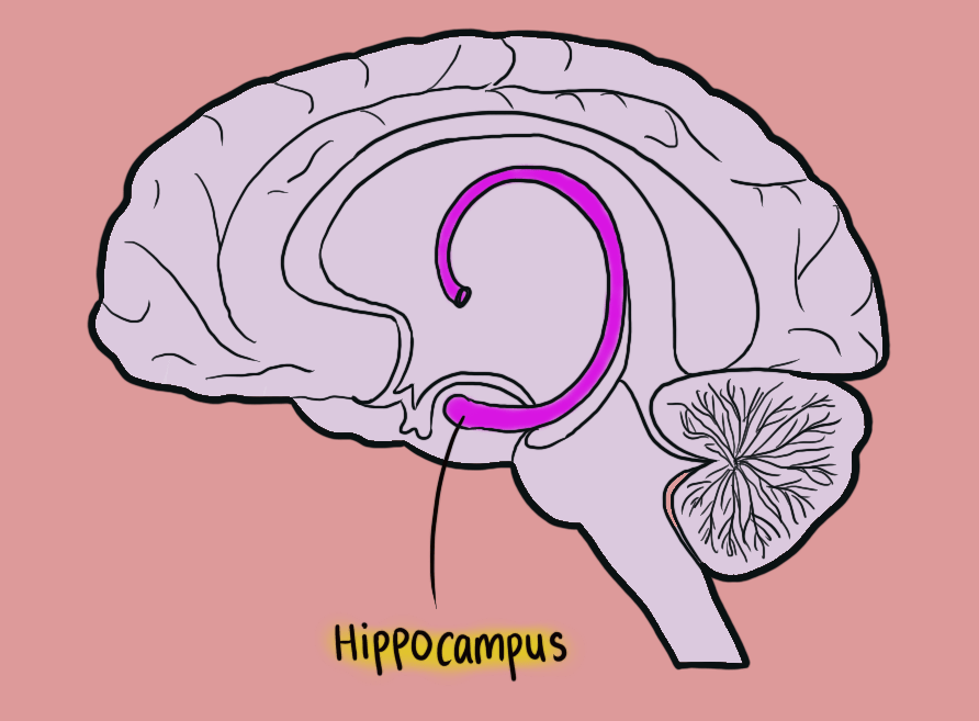
“The regions that we’re interested in during the encoding phase of this task are the regions that are sensitive to Alzheimer’s disease progression,” Brown details, “So we’re mainly looking at the hippocampus and the frontal lobes [of the brain].” According to her, throughout the progression of Alzheimer’s disease, there’s a decline in hippocampal activation (blood flow to the hippocampus) during learning of the face-name pairs, and during healthy aging, there is a decline in frontal lobe activation. This is why Brown is especially interested in assessing the hippocampus and frontal lobes of the brain. “Because we’re working with a population of women who is at increased risk for developing Alzheimer’s disease, my predictions were that women with an oophorectomy would show less hippocampal activation during learning, compared to women with an oophorectomy who were taking estradiol-based hormone therapy, with the idea being that estradiol could have a role in maintaining function in this region. This prediction is being supported by our most recent findings.” Based on Brown’s region of interest analyses (which is where she and her colleagues hone in on one part of the brain at a time) women with the bilateral-salpingo oophorectomy surgery show decreased activation (blow flow) in the hippocampus. They also are seeing this in spontaneously menopausal or naturally menopausal women. Interestingly, in the group of women who have solely undergone the bilateral-salpingo oophorectomy surgery, Brown and her team saw less activation in the frontal regions of the brain, which is actually not always seen throughout the progression of Alzheimer’s disease. “With Alzheimer’s disease progression, compensatory increases in frontal lobe activation are often seen,” she says. “So our findings could be related to accelerated aging, because we usually see a decline in frontal activation throughout aging.”

After seeing these results, Brown has an idea of how her participants’ associative memory has been affected by either surgical or natural menopause, but her job is far from done. She needs to check for the quality of the images, correct for motion, smooth the images spatially to improve sensitivity to specific signals from the data, transform the images into standard anatomical space, and remove psychological noise (e.g. heart rate-related movement). “Currently, I am using the partial least squares methods to analyze the data. Partial lease squares involves assessing activation patterns across the whole brain without worrying about issues of multiple comparisons or highly-related data (like functional data).” Brown explains, “It’s cool to know that this type of statistical analysis exists and can help us answer research questions in a more sensitive and nuanced way.”

Early Biomarkers for Alzheimer’s Disease in Menopausal Women
According to Brown, the face-name task has often been presented as a way of looking for potential early biomarkers for Alzheimer’s Disease. “When you do these group comparisons, there are fairly consistent patterns of brain activation suggestive of a prodromal Alzheimer’s disease state,” she says. “But I would say to take this information with caution. Most researchers comparing normal aging to early Alzheimer’s Disease include women and men in the same groups. It’s challenging when you’re studying Alzheimer’s disease since you may have a limited sample size. Still, when you’re always integrating women and men, it’s possible that some critical differences are being glossed over because you’ve included people who may be showing opposite patterns of brain activation. You’ll lose some nuance,” she explains. Continuing, Brown elaborates, “There are sex differences in functional brain activation between men and women during associative encoding. There could even be differences based on menopause type – whether you’ve undergone surgical menopause or natural/spontaneous menopause.” Apart from the type of menopause a woman is going through, she also details the other factors that affect the results of this task, such as the medications someone is taking, or even the type of hormone therapy they’ve been administered.
Ultimately, while fMRI studies tasks such as the face-name task could be used as a biomarker for Alzheimer’s Disease, doctors and researchers must be careful to consider all these individual factors before drawing conclusions. “Something that I think the neuroscience community should be focusing on more, especially if women who’ve undergone menopause are included in a cohort, is asking these kinds of questions,” Brown states. “Because at the end of the day, it could have a really big influence over the results we are seeing, especially in the hippocampus. It will only serve to improve the conclusions we draw.”
